Objectives of the service
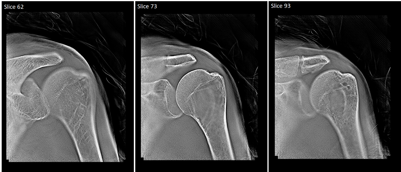
Today, 3D Computerized Tomography (CT) scanners give better results than 2D X-rays but they are too expensive and give too high a radiation dose to be used on many patients when they first present with a suspected fracture, cancer or lung disease. This means that serious conditions can be misdiagnosed leading to delayed healing of fractures, potential litigation for missed fractures, and delayed diagnosis of cancer. Adaptix’s vision is to transform radiology by fundamentally changing how X-rays are generated. Currently X-rays are still made using vacuum tubes which are big and heavy. Meanwhile, TVs have moved from vacuum tubes to flat panel displays. Adaptix‘s technology will make a similar transition for X-rays. Using an array of miniature X-ray sources that can be fired in a sequence enables 3D images to be derived from a stationary flat panel that is light enough to be brought to the patient. This means that patients can get:
-
more informative 3D imaging when they first arrive in hospital thereby saving time and cost and
-
regular low-dose 3D imaging at the bedside for longer-term conditions like lung disease when regular monitoring is needed.
Users and their needs
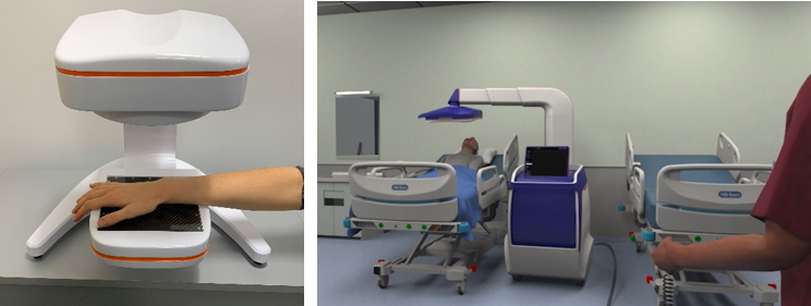
Medical imaging is needed throughout the world. The products developed through the TransDIm project can bring the following benefits:
Hospital outpatient radiology departments:
-
More sensitive detection of fractures
-
Better assessment of healing
-
Earlier detection of diseases such as lung cancer
-
Low-dose regular monitoring of conditions such as cystic fibrosis
Intensive care wards
-
Higher confidence in the positioning of tubes in ventilated patients
Podiatrists and orthopaedic surgeons
-
Ability to handle more cases on site without needing to send the patient elsewhere for a CT scan
-
Ability to perform weight-bearing imaging of the foot in the position that causes pain
Primary care & General Practice
-
Low cost device giving the option to diagnose cases in community settings avoiding the cost of sending so many patients to hospital. This could be combined with remote reading.
Developing countries and remote settings
-
Bring mobile 3D imaging in a vehicle to places that have no access to expensive scanners
Veterinarians
-
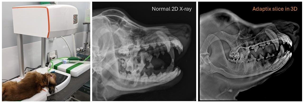
3D imaging is helpful with animals as well as humans, but most veterinary practices only have 2D X-ray as CT is too expensive for their customers. This is a separate market to human imaging but an attractive one and also allows earlier testing of products.
Service/ system concept
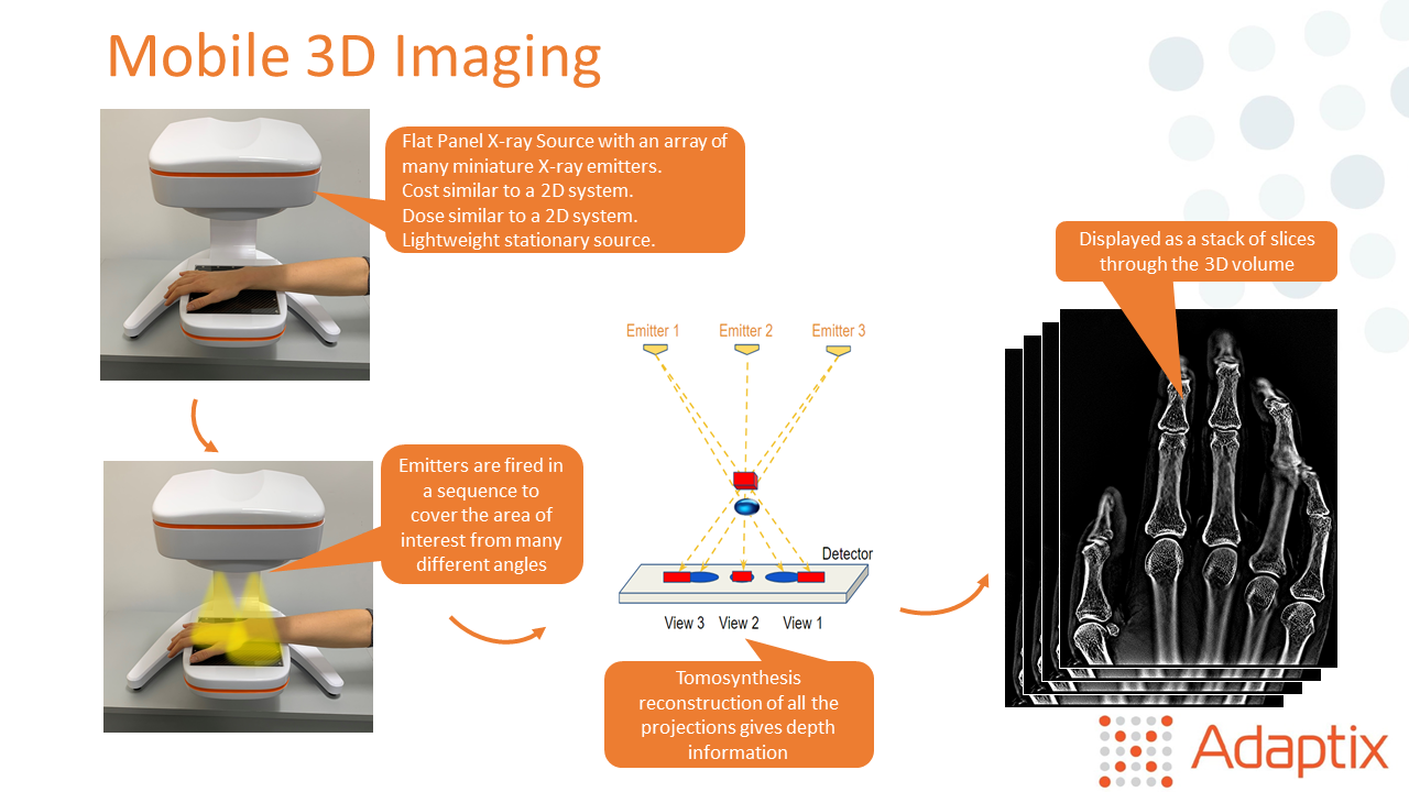
An array of miniature X-ray emitters are fired in a sequence from a novel, patented flat panel X-ray source. In the same way that having two eyes provides depth perception, firing X-rays from multiple positions allows the 3D structure of subjects to be derived, enabling this information to be displayed to the user as a stack of slices. This gives much more information than a simple 2D X-ray with overlapping bones collapsed into a single shadow.
Space Added Value
The product developed by Adaptix is using technologies derived from those initially developed for a number of Space missions, X-rays are made by firing electrons through a vacuum into metal. Conventional systems use vacuum tubes that rely on 'thermionic emission' of electrons where a cathode is heated to a high temperature to give the electrons enough energy to escape into the vacuum. A more efficient system in terms of size and power is to use 'field emission' where sharp tips on the cathode create field gradients strong enough to pull out electrons. The field emitters used in this technology have a shared heritage with some space systems. Miniature electron emitters are beneficial for a variety of purposes:
-
Ion propulsion as an energy-efficient means of providing thrust to spacecraft in the vacuum of space
-
Field neutralisation as an important means of preventing the unwanted build-up of charge
-
Ionising gases for analysis e.g. the Ptolemy device on the Philae lander of ESA's Rosetta mission, which rendezvoused with a comet in 2014. The severe constraints on the size, mass and power available for Ptolemy required the miniaturisation of every component. The gas was ionised by an electron-source using field emitters and then a counter enabled isotope ratios to be measured to very high precision.
Current Status
The original project completed its final review in March 2023 and has now been extended. During the initial project phase, Adaptix brought its first two products to market.
The first is a mobile 3D system for veterinary imaging. This compact device runs off a normal mains socket so is easy to install and use in an existing radiology room. In contrast, a CT scanner requires 3 phase power, air conditioning and special shielding. The systems have been used for imaging the teeth, heads and limbs of cats, dogs and rabbits, and also some more exotic species such as bearded dragons. Adaptix now have many commercial installations around the UK and made the first sale in the USA in 2024.
The second product is for low-cost, low-dose, mobile, orthopaedic, 3D imaging in humans. The regulatory pathway for human imaging is longer than for veterinary imaging. However, Adaptix received a 510(k) clearance by the US Food & Drug Administration in January 2023 for a first, smaller orthopaedic imaging system for imaging hands, elbows and feet. The development of a larger system that can also image knees and shoulders has now completed its on-Site Application Test ahead of starting live human imaging in clinical studies.
